AEM: The microbiome dynamics and interaction of endosymbiotic Symbiodiniaceae and fungi indicated thermal bleaching susceptibility of coral holobiont
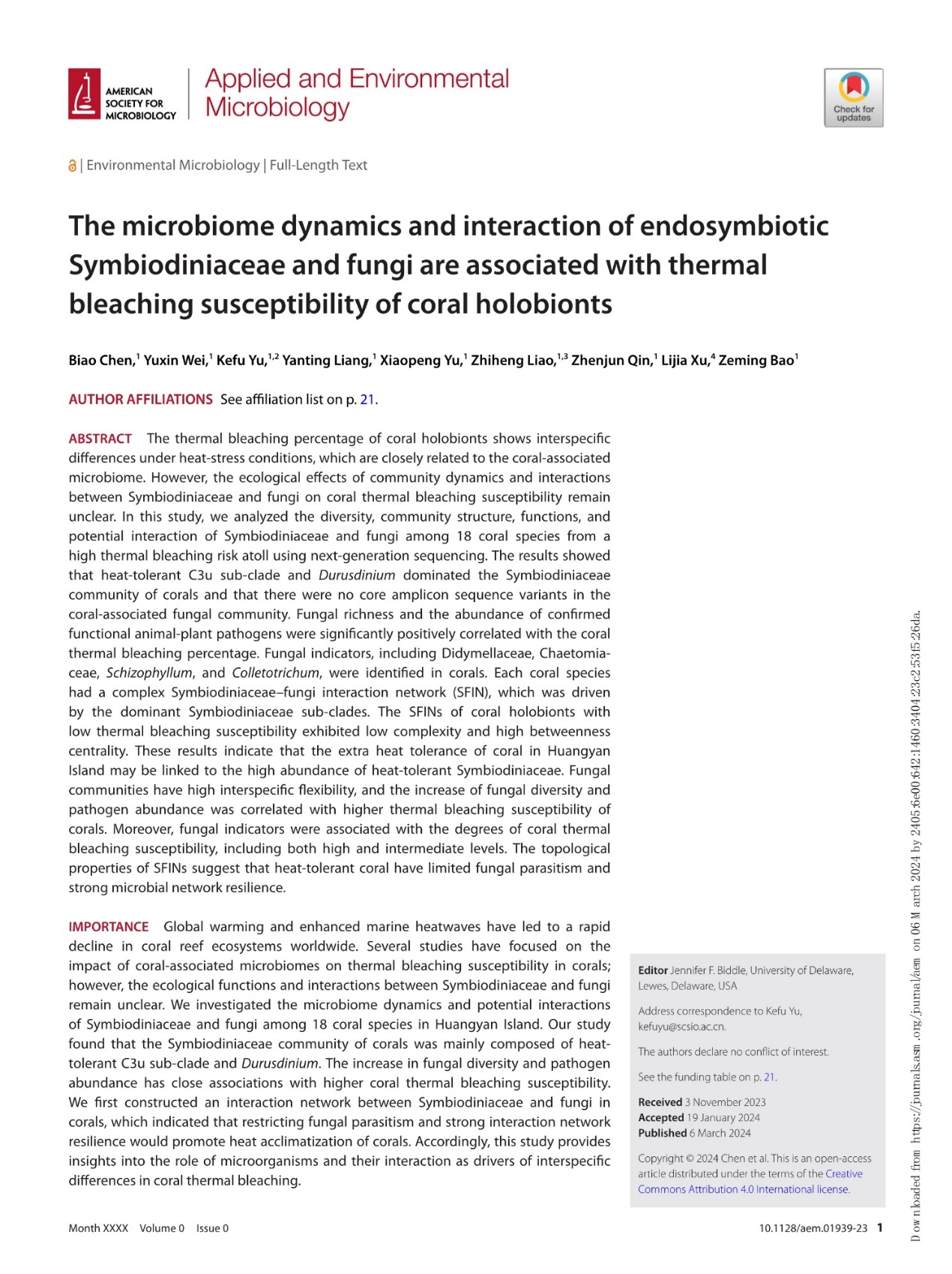
The academic article “The microbiome dynamics and interaction of endosymbiotic Symbiodiniaceae and fungi are associated with thermal bleaching susceptibility of coral holobiont” has been published in Applied and Environmental Microbiology (AEM;JCR Q1). The first author of the article is Assistant Professor Biao Chen from the Department of Marine Biology and Ecology. TheAEM is a prestigious journal published by the American Society for Microbiology.
The frequency of mass coral bleaching events has increased due to global warming, but reef-building corals exhibit significant interspecific differences in their bleaching susceptibility. Coral bleaching susceptibility is influenced not only by the coral host but also by the associated microbiome. Previous studies have primarily focused on the effects of individual microbial components on coral thermal tolerance, with fungi—an important part of the coral microbiome—being underexplored. This research gap hinders a comprehensive understanding of how the microbiome shapes variations in coral bleaching susceptibility.
This study examines 18 coral species from Huangyan Island in the South China Sea, using the bleaching percentage of corals from 15-20°N in the South China Sea during the extreme heat event of 2020 as a quantitative measure of bleaching susceptibility. It employs microbiome analysis to investigate the dynamics, functional characteristics, and interactions of coral symbiodiniaceae and fungal communities. The key findings are as follows:
The heat-tolerant symbiodiniaceae subclades C3u and Durusdinium dominate the 18 coral species at Huangyan Island, resembling the symbiodiniaceae community composition of corals in the consistently high-temperature environment of the Nansha Islands. This suggests that Huangyan Island corals may enhance their thermal adaptation by establishing stable symbioses with highly abundant thermotolerant symbiodiniaceae.
The 18 coral species at Huangyan Island do not share a core fungal microbiome, and both fungal richness and the abundance of plant and animal pathogens are significantly positively correlated with coral bleaching susceptibility. This indicates that coral fungal communities are highly flexible, and that increased diversity and pathogen abundance may weaken coral resistance to bleaching.
Within the coral fungal community, Didymellaceae, Chaetomiaceae, Schizophyllum, and Colletotrichum serve as identifiable biomarkers of bleaching susceptibility, capable of indicating both high and moderate levels of bleaching susceptibility.
All 18 coral species from Huangyan Island exhibit complex interaction networks between symbiodiniaceae and fungi. Coral species with lower bleaching susceptibility typically have less complex networks and higher betweenness centrality, suggesting that thermotolerant corals are characterized by limited fungal parasitism and greater resilience in their microbial interaction networks.
This study is the first to establish a molecular ecological network between coral symbiodiniaceae and fungi, broadening the scientific community's understanding of how the dynamics and interactions within these communities affect coral bleaching sensitivity. Additionally, it provides direct evidence of the microbial-driven mechanisms behind reef-building corals' responses and adaptations to global warming. This groundbreaking research was highlighted in an interview and report by the American Society for Microbiology (ASM) and was published on the ASM News website on March 6, 2024. The article can be accessed at: https://asm.org/Press-Releases/2024/February/Microbes-Impact-Coral-Bleaching-Susceptibility-in.
[1] Chen Biao, Wei Yuxin, Yu Kefu*, Liang Yanting, Yu Xiaopeng, Liao Zhiheng, Qin Zhenjun, Xu Lijia, Bao Zeming. (2024). The microbiome dynamics and interaction of endosymbiotic Symbiodiniaceae and fungi are associated with thermal bleaching susceptibility of coral holobionts. Applied and Environmental Microbiology. doi: 10.1128/aem.01939-23.
